Categories
Uncategorized

What Is Oscillometry?

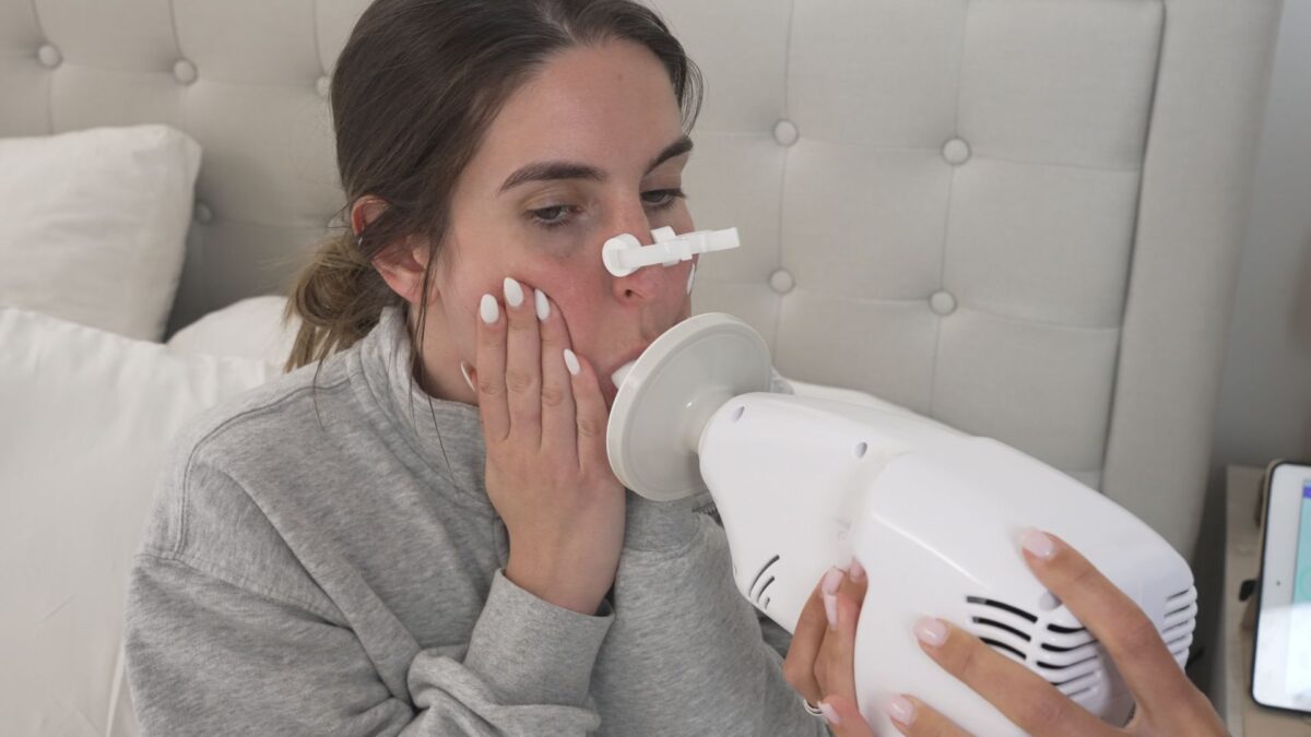
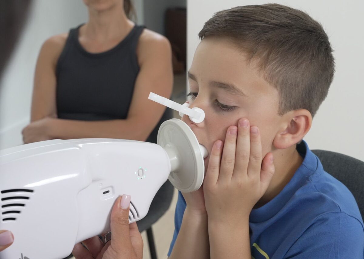
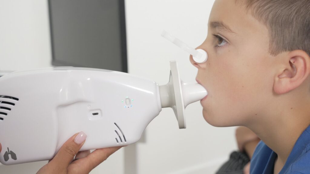
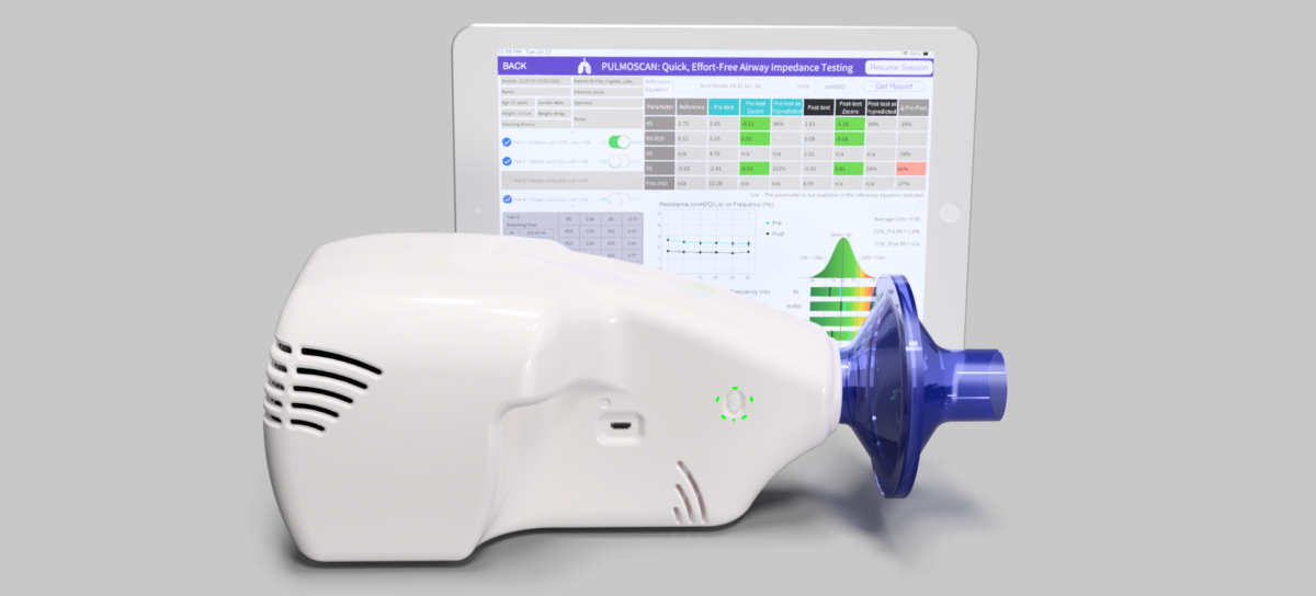
Oscillometry is a lung function test that measures how air moves through the airways during normal, quiet breathing, providing insights into obstruction and the mechanical properties of the airways.

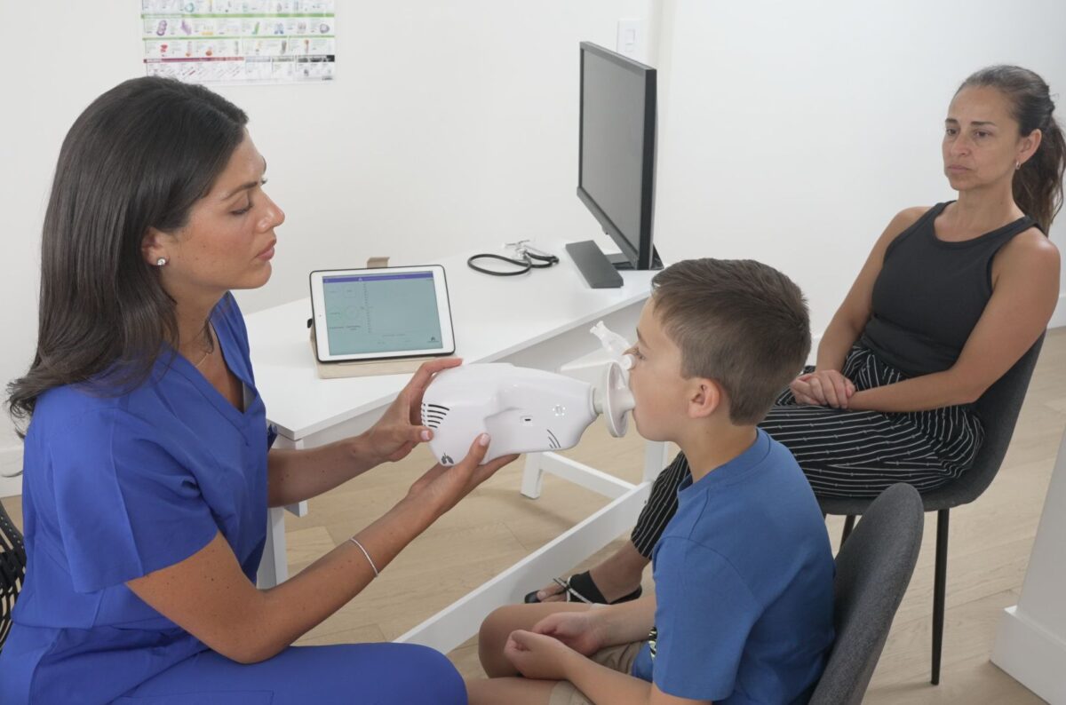
Instead of asking patients to inhale deeply or blow forcefully, oscillometry introduces mild pressure waves into the airway while the patient breathes normally. The device analyzes the resulting changes in pressure and flow due to inherent airway resistance and reactance, that is, the overall mechanical behavior of the respiratory system.
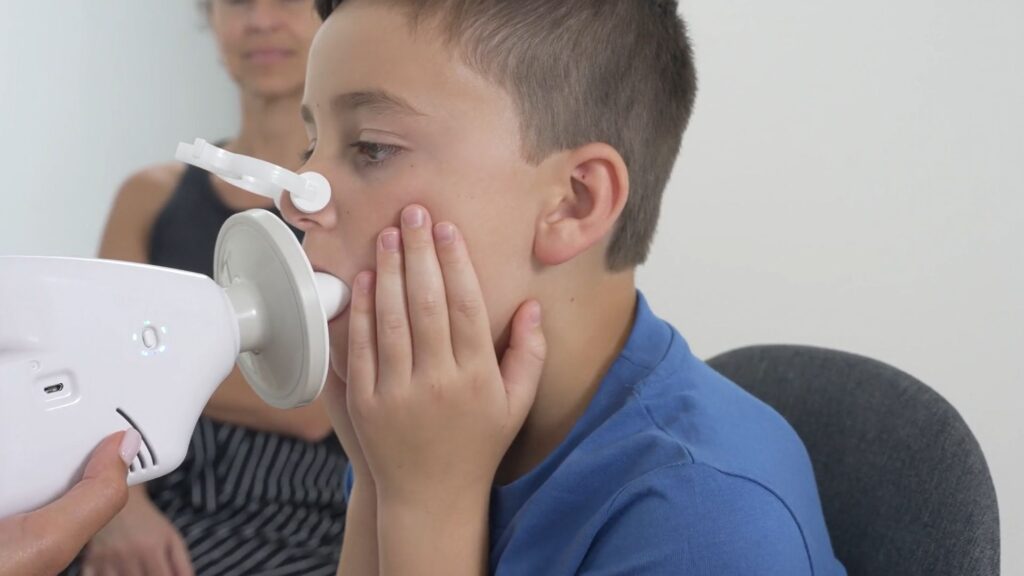
Breathing quietly is how patients actually breathe, and oscillometry capitalizes on natural breathing without relying on effort, coordination, or coaching.

The technique is often referred to as the Forced Oscillation Technique, or FOT. The word “forced” refers to the applied oscillating signal, not to patient effort. From the patient’s perspective, the test is completely passive.

Oscillometry measures resistance, which reflects how easily air flows through the airways. It also measures reactance, which reflects the elastic properties of lung tissue and airways.

Together, these parameters offer a window into the small and central airways with signatures specific for restrictive and obstructive diseases. Clinicians gain an immediate view of the region of the lungs where early disease often begins, and where symptoms may not yet appear in traditional lung test results. Learn more about data interpretation.
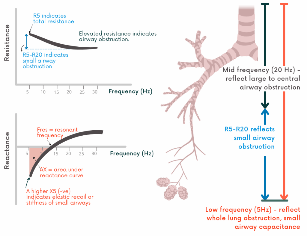

 
Oscillometry captures airway obstruction and elastic recoil, with parameters specific to small and central airway dysfunction.

Ideal for patients who cannot produce reliable forced exhalations. Children, older adults, patients with neuromuscular disease, and patients with advanced respiratory disease. 

Because oscillometry does not depend on forced maneuvers, it reduces variability caused by effort. This makes it especially useful for longitudinal tracking and for patient populations that struggle with spirometry. For vulnerable populations, oscillometry is not simply an alternative. It is often the only reliable pulmonary function testing option.

Oscillometry is already used across a range of clinical settings, including pediatric, primary care, and pulmonary practices, both internationally and within the United States. It is frequently used to support airway disease diagnosis and monitoring, particularly when spirometry is normal, inconclusive, or difficult for patients to perform.

Oscillometry is gaining popularity in pediatrics, where patient cooperation limits traditional testing. It is widely used in research to quantify airway behavior with high sensitivity, and in pharmaceutical development to evaluate treatment response, disease progression, and drug performance in both clinical trials and real-world settings. Oscillometry is also a natural fit for ER settings, where quick tests on compromised patients are beneficial.
Categories
Uncategorized

How to Interpret PulmoScan Oscillometry Data

PulmoScan uses Forced Oscillation Technique (FOT) to deliver quick, effort-free measurements of airway impedance during relaxed breathing. The device analyzes how oscillating pressure waves (5-30 Hz) travel through the respiratory system, generating resistance (R) and reactance (X) curves. 

This post walks you through interpreting oscillometry’s four key parameters, within-breath analysis, and bronchodilator reversibility using a step-by-step approach adapted from the oscillometry interpretation framework in the ERS technical standards [1], Gupta et al., 2021[2], and Brashier et al., 2015[3].


At-a-Glance PulmoScan Interpretation Flow

  1. Check quality metrics (COV, coherence)
  2. Look at R5, total resistance
  3. Look at R5–R20, small airway resistance
  4. Look at X5 & AX, small airway compliance & burden
  5. Determine pattern (obstructive, peripheral, restrictive)
  6. Add within-breath analysis, EFL or IFL detection
  7. Consider reversibility, pre- and post-bronchodilator

Step 1: Quality Check Before Interpretation

  • COV (Coefficient of Variation): <10% for adults, <15% for children (R5 parameter)
  • Repeat at least 3 tests for a valid session.
  • Coherence: >0.85 ensures signal reliability

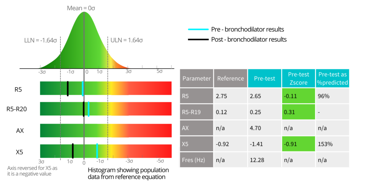
PulmoScan’s software incorporates reference equations from multiple published studies covering a range of ethnicities, enabling results to be compared against the most relevant population norms for each patient. The software displays Z-scores and percent predicted values for comparing the oscillometry test parameters, but the ERS technical standards [1] recommend using Z-scores from reference equations [4, 5]. PulmoScan software flags values beyond the upper limits of normal or ULN (1.645 Z-score) and grades severity automatically, supporting consistent, evidence-based interpretation. 

Step 2: Start with R5: Total Airway Resistance

  • R5 (resistance at 5 Hz) reflects the total resistance from central and peripheral airways.
  • ↑ R5 suggests overall airway narrowing, as in asthma, COPD, or acute bronchoconstriction.
  • Normal R5 means total airway resistance is within expected limits — but you must still check small airway parameters.

Step 3: Assess Small Airway Resistance: R5-R20

  • R20 (resistance at 20 Hz) primarily reflects large/central airways.
  • R5-R20 isolates peripheral airway resistance.
  • ↑ R5-R20 means small airway narrowing, often the earliest sign of obstructive lung disease.
  • A normal R20 but elevated R5-R20 can point to isolated small airway disease, which spirometry often misses.
Impedance parameters measured by lung oscillometry test – resistance and reactance from 5-30Hz.

 Step 4a: Look at X5: Small Airway Reactance

  • X5 (reactance at 5 Hz) reflects the elastic and inertial properties of peripheral airways.
  • More negative ↑ X5 values mean stiffer, less compliant small airways.
  • Seen in restriction (fibrosis, interstitial lung disease) and in severe small airway obstruction (asthma, COPD).

Step 4b: Review AX: Area under the Reactance curve

  • AX is the integrated area under the reactance curve from 5 Hz to Fres (resonant frequency or zero-crossing).
  • ↑ AX indicates greater small airway dysfunction and can amplify findings from X5.
  • AX is highly sensitive to early changes in the small airways.

Step 5: Identify the Pattern

Obstructive

  • ↑ R5, ↑ R5–R20, ↑ X5, ↑ AX
  • Seen in asthma, COPD, bronchiolitis
Peripheral Airway Obstruction
  • Normal- R20, ↑ R5–R20, ↑ X5, ↑ AX
  • Often early asthma/COPD
Typical pattern of oscillometry parameters observed for peripheral airway obstruction
 

Restrictive 

  • Normal/slightly ↑ R5, normal R5–R20, markedly ↑ X5, ↑ AX
  • Seen in fibrosis, ILD, hyperinflation
Typical pattern of oscillometry parameters observed for restrictive airway obstruction

Obstructive with central airway component

  • ↑ R5, ↑ R20, ↑ X5, ↑ AX
  • Normal R5–R20
  • Seen in asthma

For more disease cases, please refer to the examples provided in Gupta et al., 2021[2].

Step 6: Use Within-Breath Parameters for Deeper Insight

PulmoScan can analyze inspiratory vs. expiratory phases within each tidal breath, revealing patterns invisible to whole-breath averages. These within-breath parameters help detect flow limitation:

Expiratory Flow Limitation (EFL)

  • Defined as markedly higher expiratory resistance/reactance compared to inspiration.
  • EFL is a hallmark of COPD, severe asthma, and other obstructive diseases.
  • Mechanism: During expiration, small airways collapse prematurely, trapping air.
  • Oscillometry signal:
    • R5 (exp) markedly higher than R5 (insp)
    • More negative X5 (exp) than X5 (insp)
    • Often seen with dynamic hyperinflation.

Inspiratory Flow Limitation (IFL)

  • Characterized by higher inspiratory resistance/reactance compared to expiration.
  • Suggestive of restrictive mechanics, including upper airway restriction, interstitial lung disease, and chest wall disorders.
  • Mechanism: Increased stiffness or structural limitation reduces inspiratory compliance.
  • Oscillometry signal:
    • R5 (insp) markedly higher than R5 (exp)
    • X5 more negative during inspiration.

Why It Matters:

  • Whole-breath results may appear borderline or normal, but within-breath analysis can reveal dynamic airway behavior that guides diagnosis and therapy.
  • EFL detection can prompt earlier COPD intervention.
  • IFL patterns can push work-up toward restrictive or neuromuscular causes rather than obstructive disease.

Step 7: Bronchodilator Reversibility with PulmoScan

PulmoScan can test reversibility without repeated forced maneuvers. Per ERS guidance, a significant change is:

  • R5 ↓ by ≥40%
  • X5 ↑ by ≥50%
  • AX ↓ by ≥80%

By combining whole-breath parameters with within-breath flow limitation analysis, PulmoScan helps clinicians move beyond a simple “normal/abnormal” report and toward a nuanced, physiology-driven interpretation that can:

  • Detect early disease
  • Differentiate between obstructive and restrictive mechanics
  • Identify dynamic airway collapse or stiffness patterns
  • Track patient response over time, in the clinic or at home.o

*Disclaimer: The PulmoScan Interpretation Guide is provided for general informational purposes only. To date, there is no official interpretation guide or flow chart issued by the American Thoracic Society (ATS) or the European Respiratory Society (ERS). The framework presented herein is derived from published scientific literature as cited and does not represent an official interpretation guideline or flow chart.

Cognita Labs makes no representation or warranty as to its clinical accuracy, completeness, or applicability to individual patients. The content is subject to change as new research emerges, and while Cognita Labs will make reasonable efforts to update the guide, we cannot guarantee that it will always reflect the latest evidence or consensus.

This guide is not a substitute for professional medical judgment. Physicians and other qualified healthcare providers should base their diagnostic and treatment decisions on the complete clinical picture, including the objective airway obstruction data provided by PulmoScan and, when appropriate, additional tests or assessments.

Use of this guide is at the discretion and sole responsibility of the healthcare professional. Cognita Labs disclaims any and all liability for outcomes resulting from reliance on this information.

References:

  1. King, Gregory G., et al. “Technical standards for respiratory oscillometry.” European Respiratory Journal 55.2 (2020).
  2. Gupta, Neeraj, et al. “Oscillometry–The future of estimating pulmonary functions.” Karnataka Paediatric Journal 35.2 (2021): 79-87.
  3. Brashier, Bill, and Sundeep Salvi. “Measuring lung function using sound waves: role of the forced oscillation technique and impulse oscillometry system.” Breathe 11.1 (2015): 57-65.
  4. Gochicoa-Rangel, Laura, and Mario H. Vargas. “How best to choose an oscillometer and reference equations for your patients with asthma.” Annals of Allergy, Asthma & Immunology 134.2 (2025): 159-164.
  5. Liang, Xiaolin, et al. “Clinical application of oscillometry in respiratory diseases: an impulse oscillometry registry.” ERJ open research 8.4 (2022).
Categories
Uncategorized

Clinical Partnerships for Early Detection

Routine Lung Function Screening in Primary, Pediatric, and Pulmonary Care

Across conversations with pediatric, primary, and pulmonology care teams, one point of consensus clearly emerges: Earlier detection matters, and diagnostic tools must seamlessly integrate into existing workflows to hold practical value, especially as routine lung function screening becomes the new standard.

Cognita Labs partners with forward-thinking clinics to help them evaluate routine lung function screening as a standard part of care. These collaborations generate measurable, real-world insights – not just about the technology, but about how earlier detection improves outcomes without disrupting workflows.

Clinical studies demonstrate the need for routine lung function screens:

  • 1 in 4 patients with measurable airflow limitation had no prior diagnosis of a respiratory condition¹.
  • Screening with oscillometry detected early changes even in asymptomatic patients – changes that spirometry often missed².
  • A sub-60-second test, often completed during vitals, identified respiratory impairment in patients as young as 4³.

We’ve designed the barrier to entry for clinics to be extremely low, and the PulmoScan solution makes this shift in care delivery not only possible but simple.

Our calibration-free, sub-minute test integrates directly into vitals, requires minimal training, and delivers clear results instantly.

This empowers providers across specialties to detect respiratory impairment earlier, for treatment in-house, or specialist referral with progress monitoring between visits.

Our pilots are focused on four key areas:

  • Workflow integration – Aligning screening with vitals collection and daily throughput.
  • Clinical decision-making – Assessing how immediate results inform diagnosis and treatment for asthma, COPD, and other respiratory diseases.
  • Patient and family engagement – Evaluating how quick, non-invasive screening improves trust, adherence, and follow-up among patients and their families.
  • Implementation support – Identifying training, reporting, and follow-up processes that sustain long-term success for both patients and staff.

Participating Pilot Clinics Receive:

  • Early access to a calibration-free device with results available before the provider enters the exam room.
  • A shared data environment to inform both day-to-day care and broader care models.
  • Guidance on implementation and reimbursement.

With PulmoScan, proactive respiratory care moves from concept to clinic in days, not years. As routine lung screening becomes the norm, respiratory care is shifting from reactive to proactive care models.

We’re inviting care teams ready to set a new standard in early lung disease detection to join this effort.

Getting started is simple. Schedule a demo today through our quick contact form, or reach out directly at ([email protected]) to discuss how the program can fit your workflow and clinic.

Sources

  1. Sinyor, M., et al. (2022). Under- and over-diagnosis of asthma: a global review. Respiratory Research, 23(1), 241. https://respiratory-research.biomedcentral.com/articles/10.1186/s12931-022-02170-y
  2. Garg, P., et al. (2020). Spirometry vs Impulse oscillometry in evaluation of children with asthma. European Respiratory Journal, 56(suppl 64), 4252. https://publications.ersnet.org/content/erj/56/suppl64/2647
  3. PulmoScan Website. PulmoScan: Oscillometry for Pediatric Lung Function Testing. Cognita Labs. https://pulmoscan.cognitalabs.com/
Categories
Uncategorized

Hidden in Plain Sight: Why Lung Function Decline Gets Missed

The Patient Presents as Healthy Overall – But Decline Starts Quietly

A child who gets winded too quickly. An adult who underreports shortness of breath. A lingering cough that doesn’t meet formal criteria for referral. This is how early lung function decline shows up – not as a loud siren, but often as a whisper.

In pediatrics, primary care, and even asthma clinics, declining lung health often evades detection. Symptoms emerge slowly. Patients downplay them. And clinicians lean toward watchful waiting – not out of neglect, but because the tools to investigate those suspicions don’t fit the realities of daily care.

Our Tools Just Don’t Fit Clinicians know lung health matters. But spirometry asks a lot:

  • Expert coaching from
    Routine spirometry can be challenging for most patients, particularly our vulnerable patients – children, elderly, and high disease severity.
    trained respiratory therapists
  • Multiple repeated efforts
  • Extra training and time
  • Poor fit for younger or harder-to-test patients

Many teams don’t have that capacity in-house. And even when performed correctly, most spirometry tests miss small-airway decline – the type that creeps in quietly, without pronounced symptoms.

So the test gets skipped or tossed out. The condition gets missed. Not because the clinician didn’t care – but because the available tools weren’t built for routine care environments.

Why Early Respiratory Screening Matters

  • Early lung function decline is subtle, but clinically serious
  • Patients normalize symptoms until the disease has progressed
  • Delayed diagnosis means delayed treatment, worse outcomes, and higher costs
  • Over 60% of adults with moderate obstruction report no known respiratory diagnosis

The problem isn’t awareness – it’s the lack of a tool that fits the rhythm of care.

PulmoScan was designed keeping patients of all age groups and abilities in mind, tailored for busy clinics that need lung function tests performed in under a minute.

Let’s Rethink What’s Possible Together

PulmoScan was designed to answer a clinical need, and it’s constantly refined through input from primary care teams, pediatric clinics, asthma specialists; and every nurse, MA, and patient who’s shaped the workflow along the way.

This isn’t a new tool. But it is redefining the standard for respiratory care – built on clinically validated technology – and designed for practical routine care and real workflows.

PulmoScan doesn’t replace spirometry – it compliments and modernizes it. By streamlining lung assessments, even in challenging populations, it helps teams catch decline earlier and act faster.

Ready to See It in Action?

PulmoScan is already helping teams catch what current tools and workflows miss. If you’re curious how oscillometry could fit into your workflow, please request a demo. We’d love to learn what early detection might look like in your practice.

Find us on LinkedIn and join the conversation.

Categories
Uncategorized

Rethinking Lung Function Tests as Part of Clinic Workflows

In most clinics, taking vitals sets a natural rhythm for the day – quick, routine, and sometimes a bit rushed. But that key moment with the patient – blood pressure, temperature, pulse – is a golden opportunity for something more.

What if vitals also included a fast, reliable lung function test? One that fit seamlessly into your clinic’s existing vitals workflow – without slowing it down?

We’ve spent years listening to clinic staff: Providers, nurses, medical assistants, and front desk professionals. Their wish list is clear:

  • “If it’s not fast, it won’t happen.”
  • “We need something that fits into vitals – not something that adds another step.”
  • “The data has to be meaningful, easy to interpret, and actionable for early detection.”

The PulmoScan lung function test was built with your clinic’s process in mind.

 It’s not just another piece of equipment – it’s part of a seamless workflow fit:

  • Under a minute to screen
  • No calibration, no prep, no extra room
  • Simple to learn for staff, straightforward for providers to interpret
  • Instant results – ready before the provider enters the exam room

This isn’t about adding more to your day – it’s about making what you already do even more powerful. When lung screening becomes part of vitals, early detection becomes routine. That changes everything: From asthma detection and management, to COPD risk reduction, to building patient trust. And yes – it’s a reimbursable test, making it both clinically and financially smart.

The most effective changes in healthcare aren’t always the biggest or most complex. Sometimes innovations fit into place without disrupting a single step – while transforming patient outcomes. PulmoScan makes lung function testing a natural extension of vitals, turning a daily routine into a powerful tool for early detection and better patient health.

That’s the point, after all: Making ordinary moments of care quietly life-changing.